

When you are looking into hearing devices, it is really important to keep in mind long term implications for your life. One of these important considerations is MRIs and other head scans.
If you have been diagnosed with an acoustic neuroma, often you have years of MRI follow ups. It seems pretty standard to have 10 years of MRIs after surgery for an acoustic neuroma. If someone is doing watch and wait or radiation, there may be even more years of scans. Being able to follow and check for tumor growth or regrowth is really important.
I often get asked if there are difficulties or discomforts in doing an MRI with a bone anchored hearing device. I have had many MRIs in the ten years I have had my device, and have never had any discomfort. I do always make sure to show the MRI card my my hearing device company gave me when I am at the appointment, to make sure the MRI specifications are safe.

Regarding difficulties, there are currently essentially two category options on the market for bone anchored hearing devices. These two options have very differing implications for MRIs and scans.
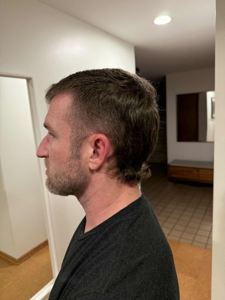
Option one is the abutment style of bone anchored devices. In this type, there is a post implanted in the skull. This is a minimally invasive procedure, and can be done awake or asleep. The post is in the skull and also is outside the skin. The skin around the post does need to be maintained in the long term, rather like a fussy piercing (regular cleaning and some sensitivity). The actual hearing device snaps onto the end of the post, which does require some finger dexterity. The hearing device itself does the vibration. Current models on the market include the Oticon Ponto and Cochlear Baha.

(my hair is held aside for visibility)

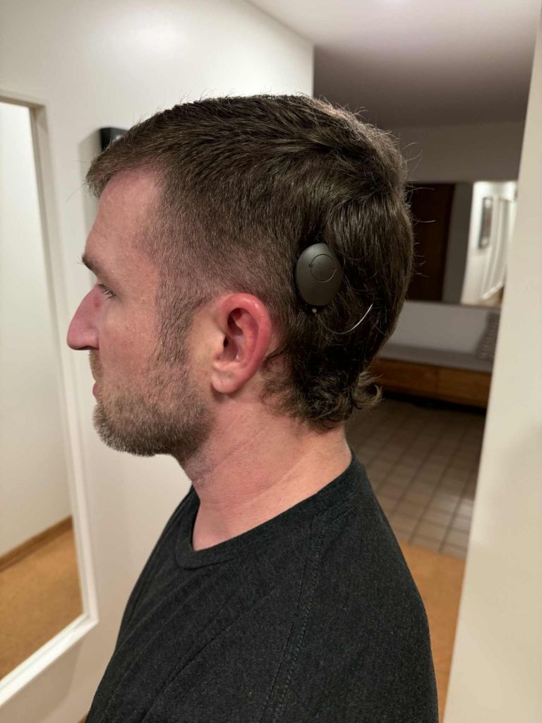
(hair moved aside for visibility)
The second option is to do a bone anchored device that is implanted. In this style, the actual vibrating device is implanted under the skin. This requires a bit of skull shaving to create the space to implant the device. There is still an external portion to the device that needs to be worn, but it attaches with a magnet. The skin can fully heal over the device with nothing coming through. It also takes much less dexterity to put on as it is just a magnet. Current models on the market include the Oticon Sentio, Cochlear Osia, and Med-el Bonebridge Samba 2.
I am not intimately acquainted with the details of every device, but I do know that the technology for the Oticon Ponto (abutment style) and the Oticon Sentio (implant) are the same. They both provide the same hearing benefits. There isn’t one that is superior to the other in technology, it just differs in attachment.
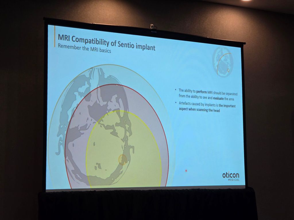
However, these styles differ hugely when it comes to MRIs. Oticon informed me that you cannot get an MRI image of the brain if you have a Sentio implant but you can with a Ponto abutment. Oticon does not recommend a Sentio if you have had an acoustic neuroma. This graph shows you the shadow the different devices cast on an MRI. The gray outer shadow shows the Sentio shadow (essentially the whole brain). The Ponto shadow is the tiniest orange circle on the graph.
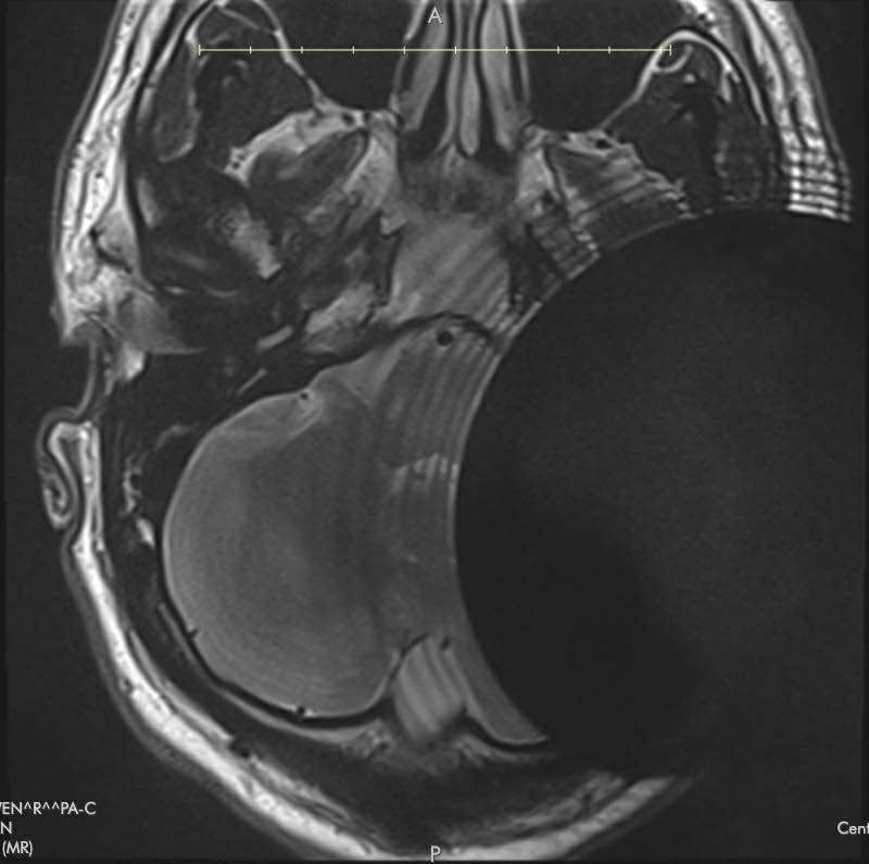
Recently, John reached out to me to share his story. John had lost hearing in one ear from an acoustic neuroma. He had chosen to get a Med-el Bonebridge implant bone anchored hearing device. His ENT doctor had told him it would cast a 17mm shadow for an MRI. However, he had a follow up MRI and it was useless for montioring his tumor. He then learned the literature from Med-el showed the shadow cast from a Bonebridge was 15cm. This is the MRI image he recieved.
He then proceeded to have a CT scan, which was not much better.
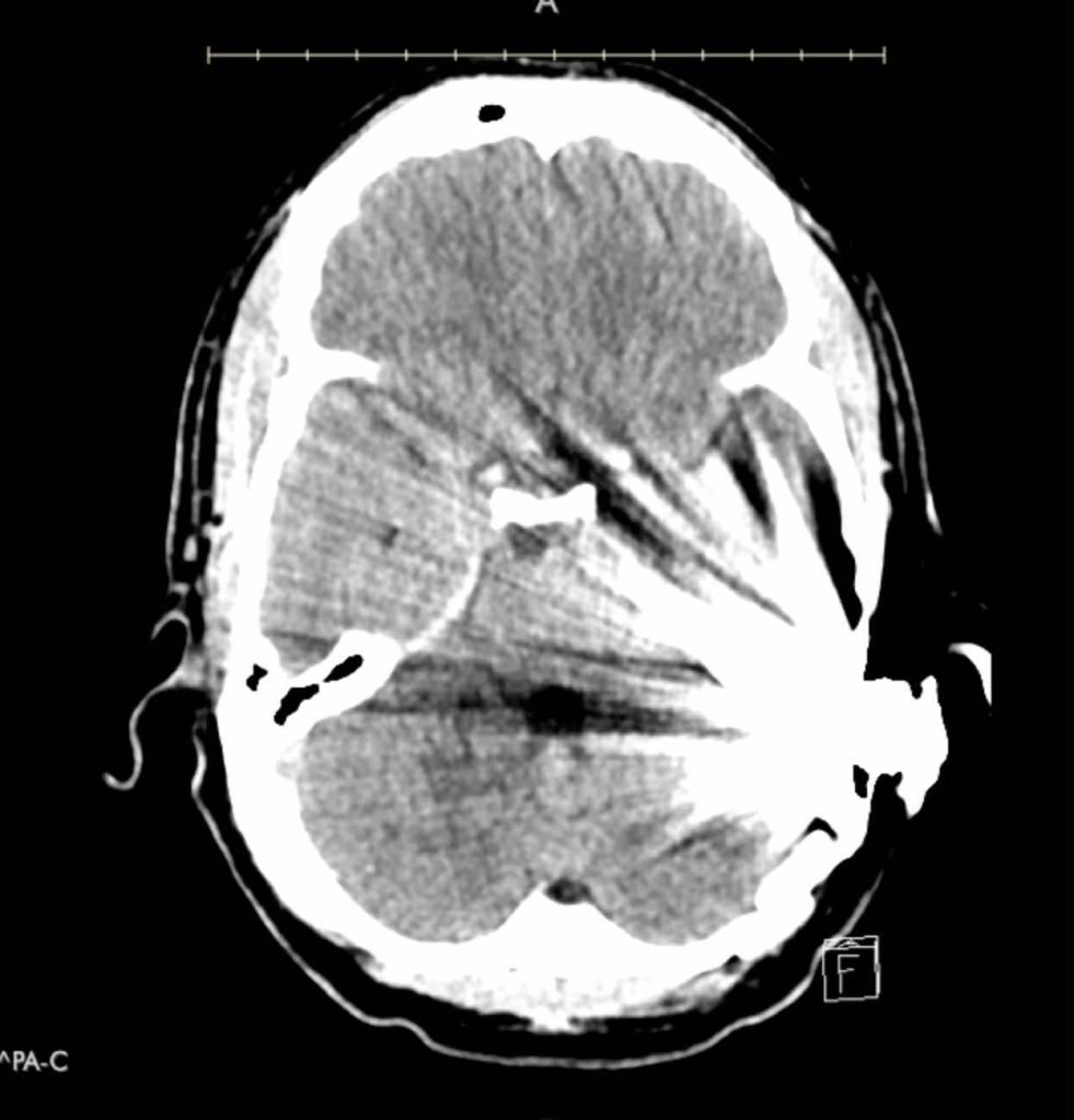
This is so very frustrating, to find out after the fact that the hearing device you have implanted prohibits you from monitoring your brain tumor.
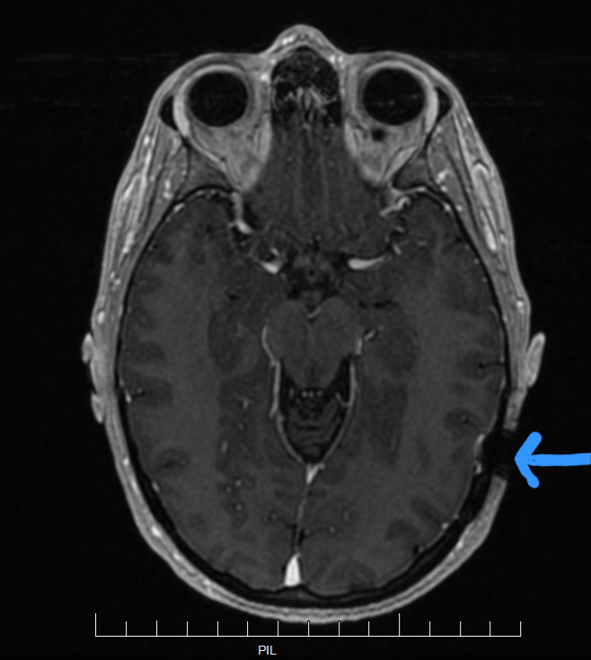
In contrast, this is my most recent MRI. The shadow from my Ponto abutment is the small black circle in the lower right side of the image. It definitely does not stop me from getting useful data from my MRIs.
I would definitely shy away from getting an implanted style bone anchored device if I believed regular brain MRIs were important for my health, such as montioring brain tumor growth. At minimum, I definitely recommend searching to find out MRI implications for any device you are considering. I think John’s story is really unfortunate. The doctors and hearing device companies should be forthright about the pros and cons of these devices.
If you would like to reach out to John, he would be happy to connect and share more about his experience: johnjreichert@gmail.com.
MRIs are definitely a possibility with a bone anchored hearing device, but which device you choose makes a huge difference on the feasibility of an MRI scan of the brain.

Leave a comment